Tag Archives: ForBio

ForBio Annual Meeting 2023 at the University Museum of Bergen

ForBio – Research School in Biosystematics held its 12th Annual Meeting at the University Museum of Bergen on September 19-21st.Sixty participants from Norway, Sweden, Iceland, Czech Republic, Poland, Germany and Peru presented their research results in various fields of biosystematics.

The main day of the meeting took place in the Tårnsalen – a beautiful venue at the top of the museum building and was opened by a talk by Endre Willassen on the history of marine research at UiB.

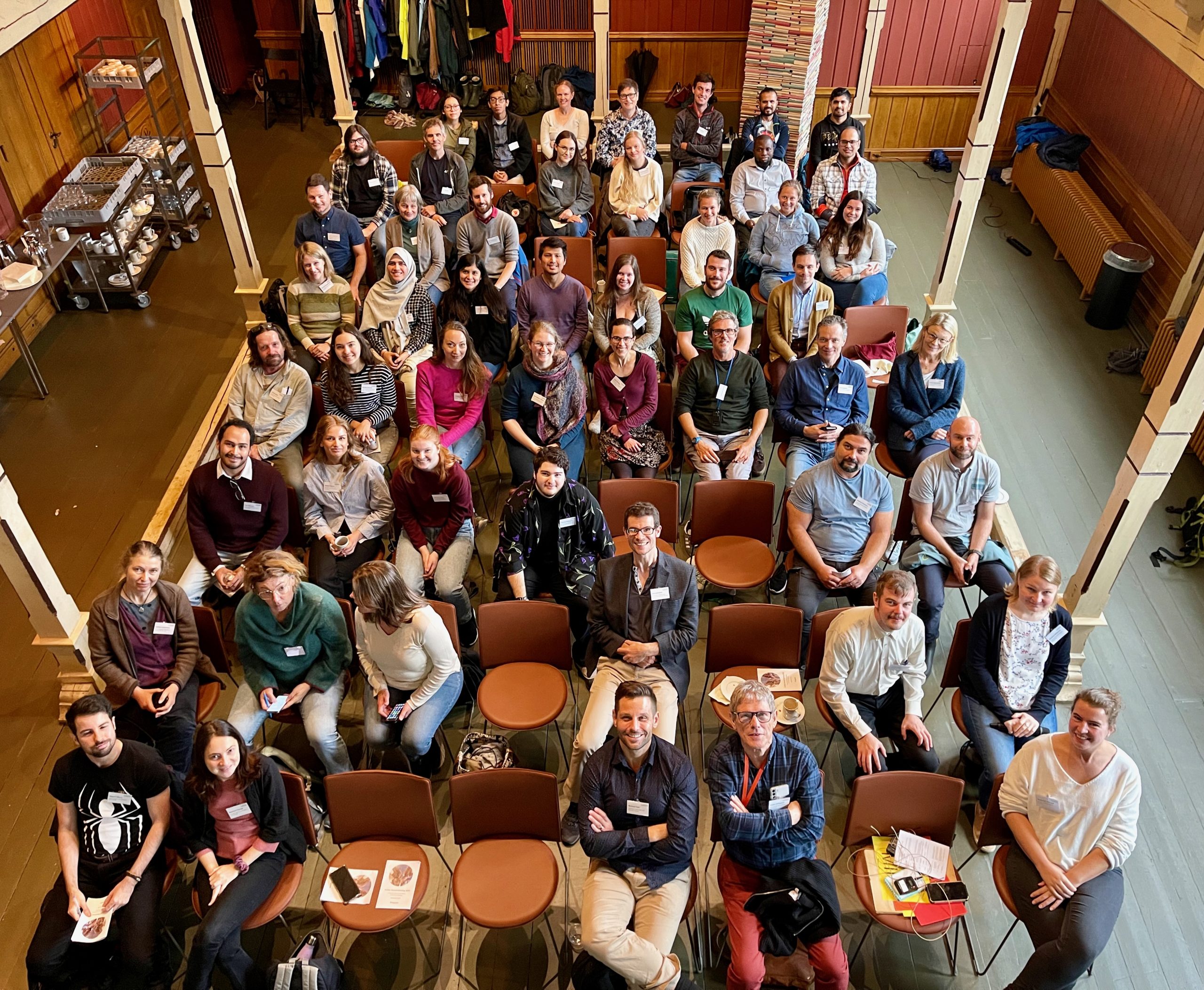
The participants - 60 of them - in the historical Tårnsalen. The photo is taken from up on a gallery in the room, so it's a bird's eye view

The participants in Tårnsalen. Photo: ForBio

Four keynote speakers gave lectures on general biodiversity research topics of high interest to a wide audience.
Michael Bok from Lund University talked about the evolution of visual systems in marine annelids. Lovisa Gustafsson from the Stavanger Botanic Garden talked about the evolution of reproductive isolation in Arctic plants. Tomas Roslin from the Swedish University of Agricultural Sciences discussed the ways to assess enormous diversity of insects using DNA barcoding, and Agnes Weiner from NORCE gave a lecture on using ancient DNA in the past marine biodiversity assessments.

ForBio [LINK to homepage] is a cooperation between the main Natural History Museums in Norway: Natural History Museum, UiO; University Museum of Berge, UiB; NTNU University Museum; and the Arctic University Museum of Norway, UiT and supported by the Norwegian Biodiversity Information Centre.

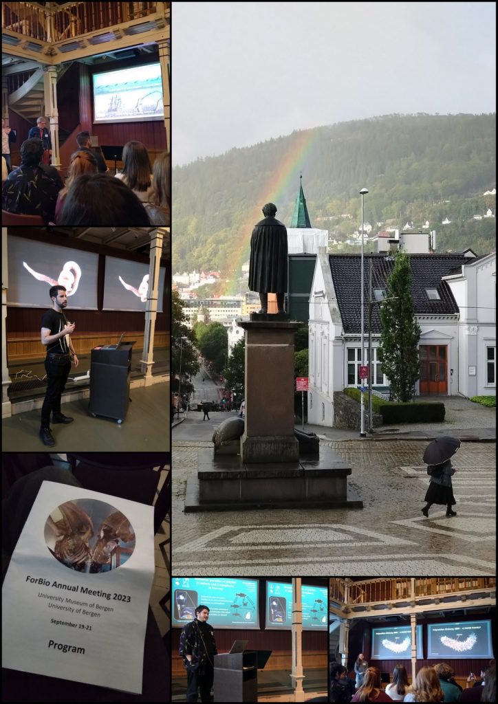
Collage showing people presenting, and a statue of Christie with a rainbow over it.

From top left: Endre Willassen presenting his talk on the history of marine research at UiB, Christie (the statue) standing outside the Museum in quite typical Bergen weather, Miguel Meca on the systematic revision of orbiniid polychaetes, the result of his PhD project, a snapshot of the program, Vincent McDaniel gives his speed talk about the parasites in jellyfishes (ParaZoo project), and Martha Everett gives her talk on the diversity of scale worms in the Arctic, the results of her PhD project

ForBio aims to advance biosystematics education for students and postdocs working in the fields of botany and zoology. The annual meetings is a great arena for the students to meet their peers and mor senior scientists and to present their research project results, exchange ideas and expand their networks.

You can find upcoming courses and events here: link

-Nataliya Budaeva
ForBio-coordinator

Hydrozoa course 2022 edition – as told by our MSc student Ana González

Last month, our project NorHydro (together with ForBio Research School of Biosystematics and project MEDUSA) organized a course on diversity, systematics and biology of Hydrozoa at the Marine Biological Station in Espegrend. Fifteen participants from 9 different countries came all the way to Bergen to learn more about these intriguing animals, share their ideas and projects, and start new collaborations. We asked one of the youngest members of the group –our highly motivated student Ana González– to share with us her thoughts about the course and her experiences with her MSc project. This is what she had to say:

When I started my Master’s Degree of Marine Ecology at the University of the Balearic IslandsI already knew about the existence of hydrozoans, but I had no idea how interesting these animals actually were. After some discussions, a lot of reading, and a fair amount of looking at pictures of hydroids and hydromedusae, I decided to work with these inconspicuous invertebrates for my MSc project under the supervision of Dr Luis Martell (University Museum of Bergen) and Dr. Maria Capa (University of the Balearic Islands). My project aims to evaluate whether we can use the benthic communities of hydrozoans as bioindicators of anthropogenic impact on the easternmost coasts of Mallorca Island, in the Mediterranean Sea.

Me on a sampling day looking for benthic hydrozoans at the marine reserve of Cala Gat (top). A closer view of the hard substrates I sample in the marine reserve (bottom left). The common hydroid Monotheca obliqua growing on Posidonia oceanica (bottom right). Picture credits: Maria Capa and Ana González.

Coastal areas are an attractive place to live, and these habitats provide ecosystem services that contribute greatly to the economy of the world, but a bad management of them can generate important damages and drastic changes in the ecosystem. One way to monitor environmental impacts in these habitats is by observing the response of their biological communities, so for this project I decided to study the assemblages of benthic hydrozoans in two opposite sites with different levels of anthropogenic impact: a harbor and a marine reserve. Moreover, I am comparing the communities in different seasons of the year, and I will analyze the assemblages growing on hard substrates (like rocks) and also those growing on a very important Mediterranean soft substrate: the endemic seagrass Posidonia oceanica.

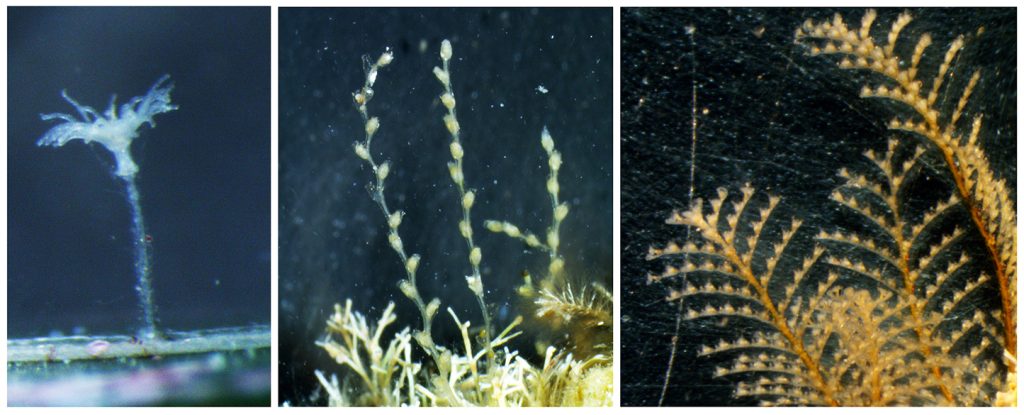
Some hydroids common in my study area are those belonging to genera Clytia (family Campanulariidae, left), Sertularella (family Sertularellidae, middle), and Aglaophenia (family Aglaopheniidae, right). Picture credits: Ana González.

At the beginning, working with benthic hydrozoans was very challenging for me since the specimens I find are easily overlooked if one is not searching carefully for them. But the more time I dedicate to observe these organisms, the more curious I became about their identity and dynamics, and the easier it was to recognize them in the samples. However, identifying hydrozoans is a difficult task and I realized early that I needed some help, so I was very happy when the opportunity arose to apply for the course “Diversity, Systematics and Biology of Hydrozoa” in Bergen. There, I had the chance to meet some of the leading scientific experts in the field that helped me understand better the taxonomy and ecology of these animals. I couldn’t have imagined how much I was going to learn during the different activities of the course, but at the end these organisms were able to catch my attention and time flew between lectures, sampling trips, and laboratory work. One aspect of the course that I particularly enjoyed is the fact that it brought together participants with different trajectories in science, and everybody was happy to share their experiences in the world of hydrozoan science.

We had all kinds of weather during the course: rain, sun, wind, and even snow! Picture credits: Lara Beckmann and Joan J Soto Àngel.

We had the chance to sample on board the UiB research vessel Hans Brattström and we collected several planktonic and benthic hydrozoans in the fjords around the Marine Station. After each sampling event, we went back to the lab to sort the samples, find the hydrozoans and identify them to species. The plankton samples were usually the first ones to be processed, since hydromedusae are quite fragile and they tend to suffer morphological damages after being sampled with a net. We tried to identify all specimens to species level, with the aid of the stereomicroscopes and scientific literature with identification keys that the curse provided. The benthic samples were placed in aquariums to keep the organisms alive and then each of us had the opportunity to observe the specimens in our own stereomicroscope.

A sampling day on board of RV Hans Brattström. Top left: deploying the plankton net. Top right: a full cod-end with plankton sample. Middle right: students and teachers ready to leave the pier. Bottom: benthos sampling with the triangular dredge. Picture credits: Lara Beckmann, Sabine Holst, Luis Martell

Top right and left: students and teachers at the laboratory, identifying hydrozoans. Bottom left: searching for hydromedusae and siphonophores in the plankton sample. Picture credits: Sabine Holst and Lara Beckmann.

All together, we were able to find and identify more than 40 species from all the main groups of hydrozoans, including siphonophores, trachylines, leptothecathes, and anthoathecates. Working with hydromedusae was new for me and I discovered that observing them was more challenging than identifying the polyps, but it was also interesting in its own way. The hydrozoans that caught my attention the most were the polyps from the suborder Capitata, because their morphology is very different from the hydroids that I have observed in my MSc project so far. Capitate hydroids don’t have a protective theca, they possess tentacles that end up in a ball of nematocysts (so-called capitate tentacles), and they are absent from almost all my samples from Mallorca, which are instead dominated by hydroids belonging to the Order Lepthothecata.

Top: Colony of Sarsia lovenii (Anthoathecata: Corynidae) with gonophores (i.e. reproductive buds on the polyp body). You can also see the capitate tentacles, which end in a ball of nematocysts and are typical for suborder Capitata. Bottom: Colony of Clava multicornis showing also gonophores on the polyp body, but with filiform (non-capitate) tentacles. Picture credits: Lara Beckmann (top), Joan J. Soto Àngel (bottom).

My interest for hydrozoans, the great set of experts we had as teachers, and the charismatic animals that we collected were the perfect combination for me to have an incredible experience in this course. I think that courses like these are an excellent opportunity for beginners to learn with experts from different parts of the world. Interacting with all of these amazing people was very rewarding at both cultural and scientific levels, and this whole experience motivated me to keep on studying these interesting animals that are a part of the complex functioning of our oceans.

-Ana

CT scan all the things!

Oslo Natural History Museum 02.12.2019 – 06.12.2019

Literature for the week of our CT scanning course


From December 2 till December 6 Justine Siegwald, Manuel Malaquias and I (Cessa) attended a CT scanning course in Oslo at the Natural History Museum.

The course was organized by the Research School in Biosystematics (ForBio) and Transmitting Science.

The group attending the CT scanning course in Oslo at the Natural History Museum

The three of us all work on mollusks and one of the main reasons to deepen ourselves in CT scanning techniques is because of studying the internal anatomy of these often small and delicate specimens.

Species descriptions are not only based on external morphology and/or DNA barcoding, a lot of the species differences are small details within the internal structures. Like the reproductive organs and digestive system (e.g. radular teeth) of the animals. This is very laborious work, and can be challenging especially with the smaller specimens of only a few millimeters long. Besides some species are difficult to collect and can be a rare collection item of the museum. Once you cut these animals to study them, there is no turning back and the specimen is basically destroyed. Being able to see the insides of the animal without touching it would therefore be ideal. CT scanning comes very close to that and with powerful X-ray we can almost see every detail of the insides of the animals without the need to cut them open

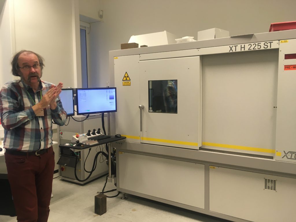
The CT scan in the Natural History Museum of Oslo with one of the teachers in front (Øyvind Hammer)

Lecture about the technology of the CT scan and X-ray

However, how easy as this sounds, CT scanning soft tissue comes with some challenges. Soft tissue means that the x-ray contrast is often very low. Even with modern good x-ray detectors it is still difficult to detect the different internal structures. Therefore, during the course, we were taught how to artificially increase the contrast of the tissue by staining the specimens before mounting them in the CT scan machine.

Coating a specimen with a metal (e.g. gold or platinum) is only useful if you want to see external details of the study object, coating will not help revealing internal structures. For radio contrasting the internal anatomy, you can stain specimens with high density liquids like iodine to enhance the x-ray contrast. So, we went for this option and left our specimens in iodine ethanol solution overnight


After the staining process, we needed the wash out any extra iodine before mounting the specimen into the CT scan. A good scan is not simply pressing the button of the machine; there are a bunch of settings that can be adjusted accordingly (e.g. tube voltage, tube current, filament current, spot size, exposure time, magnification, shading correction, etc.). The scan itself can take hours and file sizes of 24GB for a single object are not uncommon, which means you need a powerful computer with decent software to process this information. Part of the course was also about how to visualize and analyze the files of the CT scan with software like Avizo

Video 1. Software Avizo has a user-friendly interface for analyzing all sorts of CT scanning files.

The week gave us some very promising first results (se video below), and also new insights about how to increase the X-ray contrast of our sea slug samples (lots of hours of staining and very short washing steps).

Video 2. 3D reconstruction of one of Justine’s shell samples

It was a very fruitful week and besides interesting new ideas for scanning our museum specimens, we also met many old and new friends during the week that was as inspiring. ForBio and Transmitting Science did a really great job with setting up this course and I can highly recommend it to anyone who is interested in CT scanning.

Explore the world, read the invertebrate blogs!

-Cessa

Teaching DNA barcoding in Siberia

Endre, Katrine, Nataliya and Tom have recently been on a journey far removed from the ocean – although the location did hold a lot of fresh water…!

It really does look and feel a lot like an ocean…

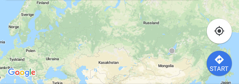
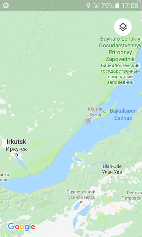
We have – together with Torbjørn Ekrem from the NTNU University Museum – been teaching DNA barcoding at the Russian-Norwegian course “Data mobilization skills: training on mobilizing biodiversity data using GBIF and BOLD tools”, which was held in Naratey on the western shore of Lake Baikal September 14-20, 2018!

The course consisted of two modules focusing on GBIF and BOLD tools. The GBIF part was taught by Dag Endresen from UiO, Laura Russell and Dmitry Schigel from the GBIF Secretariat.

It included both online preparatory work for the students and (mainly) onsite components. The online portion consisted of tasks that the students completed on GBIF’s eLearning portal.

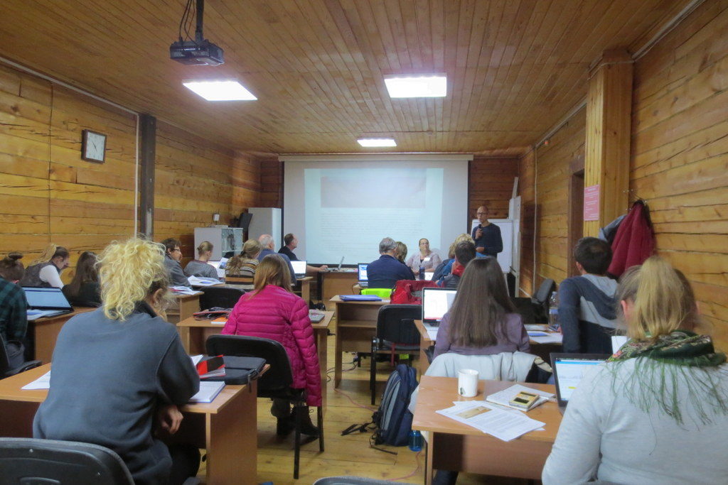
The onsite work was comprised of 20 different sessions of lectures and practical exercises, the latter with a significant component of group work.

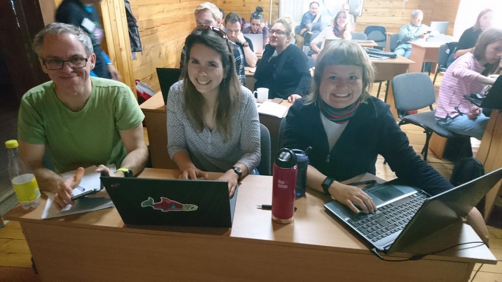
16 students from Norwegian and Russian intuitions participated, and did a wonderful job of assimilating a lot of information in a short amount of time, and turning it into practical skills.

The two main platforms we used were GBIF and BOLD – two large depositories for different kinds of biodiversity data. The GBIF-part of the course focused on the technical aspects of data mobilization, such as data capture, and management and online publishing of biodiversity data in order to increase the amount, richness and quality of data published through the GBIF network.
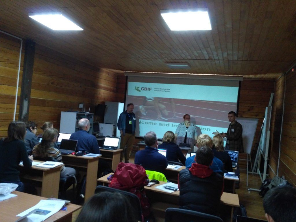
Team GBIF getting set up Photo: N. Ivanova
BOLD; Barcode of Life Data Systems

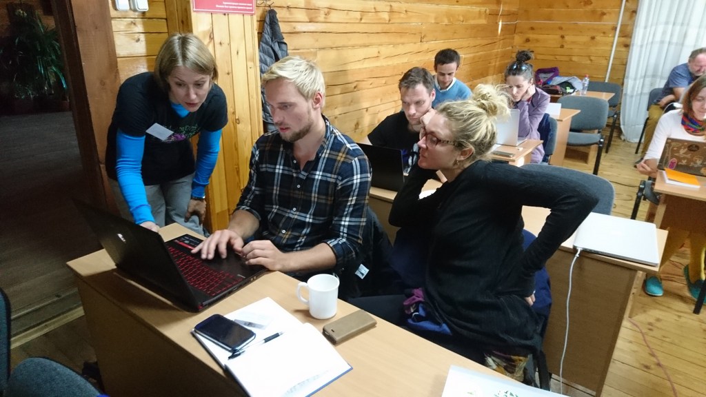
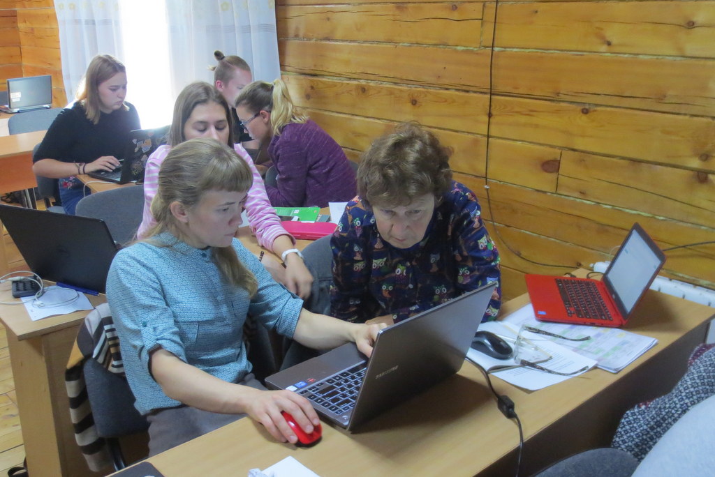
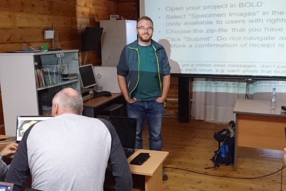
The barcoding part was aimed at both users and providers of barcoding data, and began with an introduction to the barcoding concept, and a case study of integrating data from BOLD and GBIF. This was followed by a session on the use of BOLD: creating projects and datasets, and the uploading of data, images, sequences and trace files. The students got to try all of this for themselves, and we were impressed by how well they worked together to find solutions and teach each other valuable tricks to solve the challenges.

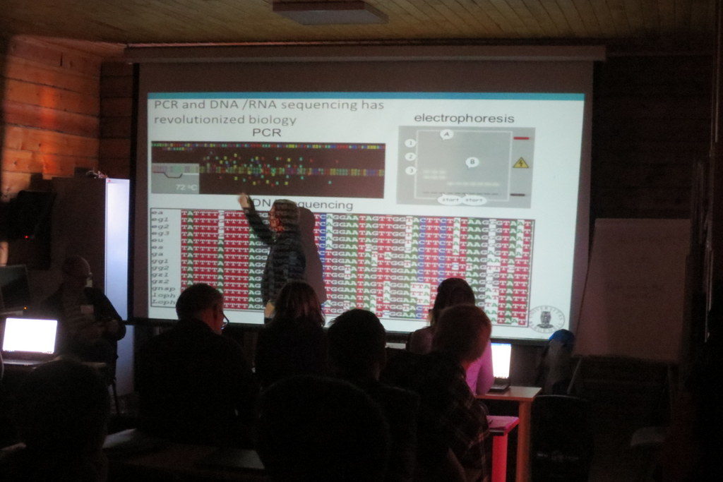
Following the lessons on how to get sequence data into the database, we covered basics of sequence analysis, and gave an introduction to the free software MEGA X which can be used for sequence alignment, translation and phylogeny.

Working in MEGA Photo: k. Kongshavn

This was again followed by a practical session in MEGA on a given data set. We also had a session presenting the analytical tools in BOLD, with a practical session exploring a dataset from NTNU. Our lessons were very well received by the students, with an average score of 4.8 out of a possible 5 on the evaluations – nice feedback for the teachers!
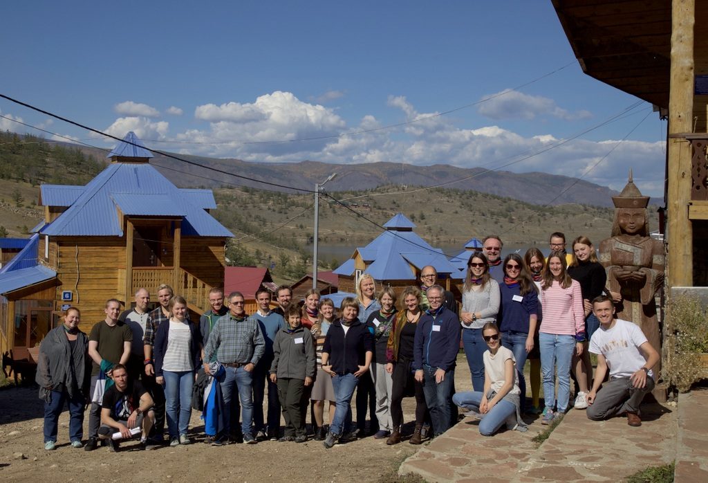
Students and teachers gathered in the Siberian sun
Photo: Dmitry Schigel, CC-BY-SA

The final task for the students was to present their presentations for “The Baikal Biodiversity Challenge”, which they were presented with on the first day of the course.
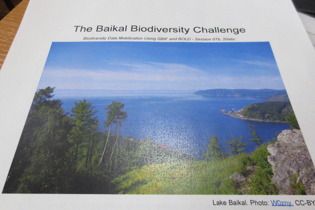
The Baikal Biodiversity Challenge

The challenge was to develop a biodiversity inventory project to map and analyze the diversity of a selected animal group. To do so they would need to use available information in BOLD, GBIF and other sources to examine what was known and identify information that was missing, and come up with suggestions on how it could be solved. It was not the easiest of tasks, however all four groups gave excellent presentations.


Group selfie wearing the NorBOL buff scarves – #mydnabarcode! Photo by Laura Russell, CC-BY-SA

The course was arranged as collaboration between the University of Bergen, the Siberian Institute of Plant Physiology and Biochemistry Russian Academy of Sciences (SIPPB SB RAS), the Global Biodiversity Information Facility (GBIF) Secretariat. NorBOL (Norwegian Barcode of Life) supplied the teachers for the barcoding part of the programme, namely Endre, Torbjørn, Katrine and Tom. Funding came from the Norwegian Centre for International Cooperation in Education (previously SIU, now DIKU), GBIF and the Research School in Biosystematics (ForBio).
For those who might not know, ForBio is a teaching and research initiative coordinated by the Natural History Museum in Oslo, the University Museum of Bergen, the Tromsø University Museum and the NTNU University Museum. It is funded by the Norwegian Taxonomy Initiative and the Research Council of Norway. The Research School offers a wide variety of both practical and theoretical courses in biosystematics, and provides a platform for facilitating teaching and research collaboration between Nordic research institutes.  The course portfolio is likely to have something of interest to offer if you work with anything related to biosystematics –and is open (and often free) to you if you are student, researcher or staff at universities, institutes and consulting companies.

Most of the ForBio courses are arranged in Norway or other Nordic countries – but this course was the second of a total six that are arranged as part of the SIU-funded MEDUSA*-project (Multidisciplinary EDUcation and reSearch in mArine biology in Norway and Russia), which is coordinated by Nataliya. The six courses are

  1. ForBio and DLN course: Comparative Morphology Methods (Trondheim, Norway 2018)
  2. Data mobilization skills: training on mobilizing biodiversity data using GBIF and BOLD tools (Siberia, Russia 2018)
  3. Zooplankton communities – taxonomy and methods (Espegrend Marine Biological Station, UiB, Norway, tentatively May 2019)
  4. Systematics, Morphology and Evolution of Marine Mollusks (Vostok Marine Research Station, Institute of Marine Biology, Vladivostok, Russia, September 2019)
  5. Systematics, Morphology and Evolution of Marine Annelids (Espegrend Marine Biological Station, UiB, Norway, tentatively June 2020)
  6. Diversity and Evolution of Meiobenthos (White Sea Biological Station, Moscow State University, Russia, September 2020)

We enjoyed the opportunity to visit such a remote locality, and to get to know the students and teachers – thank you all for making this such a wonderful experience!

Thanks also to the BOLD support team for excellent help before and during the course!

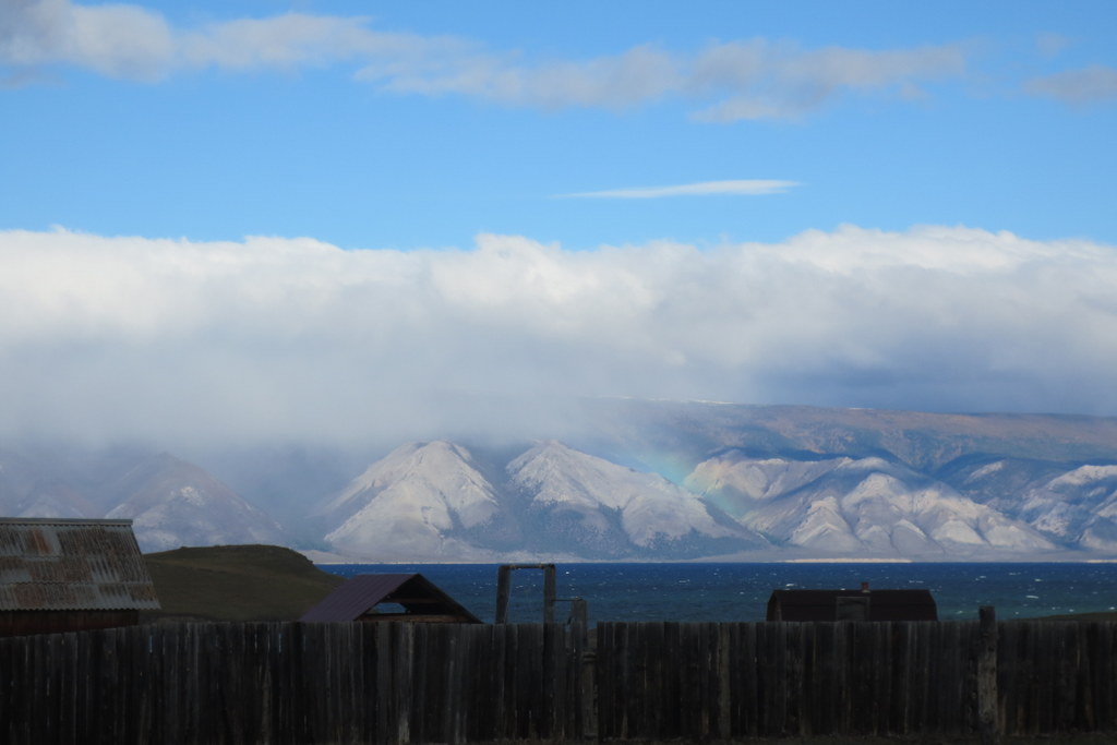
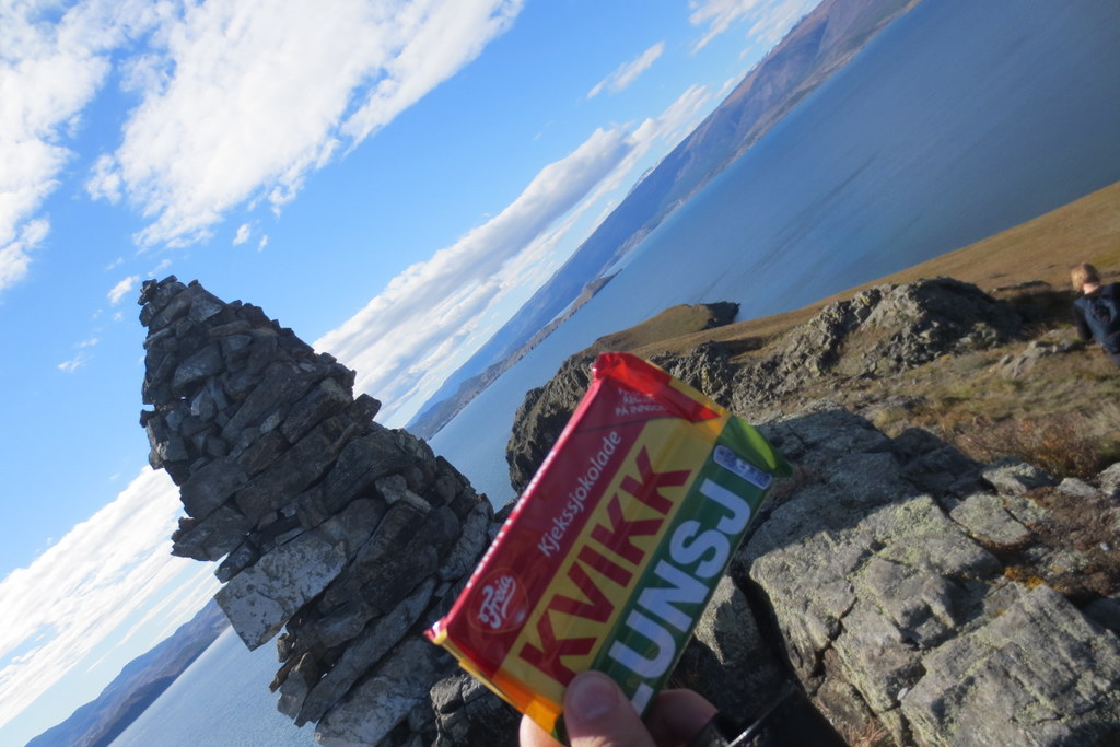
A few snapshots from the area – it was stunning! Photos by K. Kongshavn

A beautiful view from Olkhon Island (after the course), photo by K.Kongshavn

ps: we also tweeted using #ForBio_GBIF during the course
Good-bye Greenland!

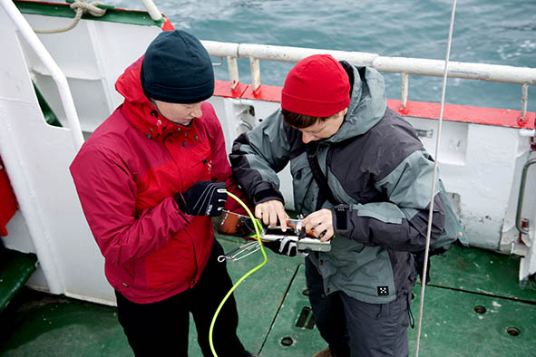
The last days before leaving the Arctic Station were busy: last boat trip, last samples, last possibility for filming work with the underwater-camera. Personal projects to finish, lab to clean, things to pack, and on top of all that: a football match against the Qeqertarsuaq “Old Boys”!

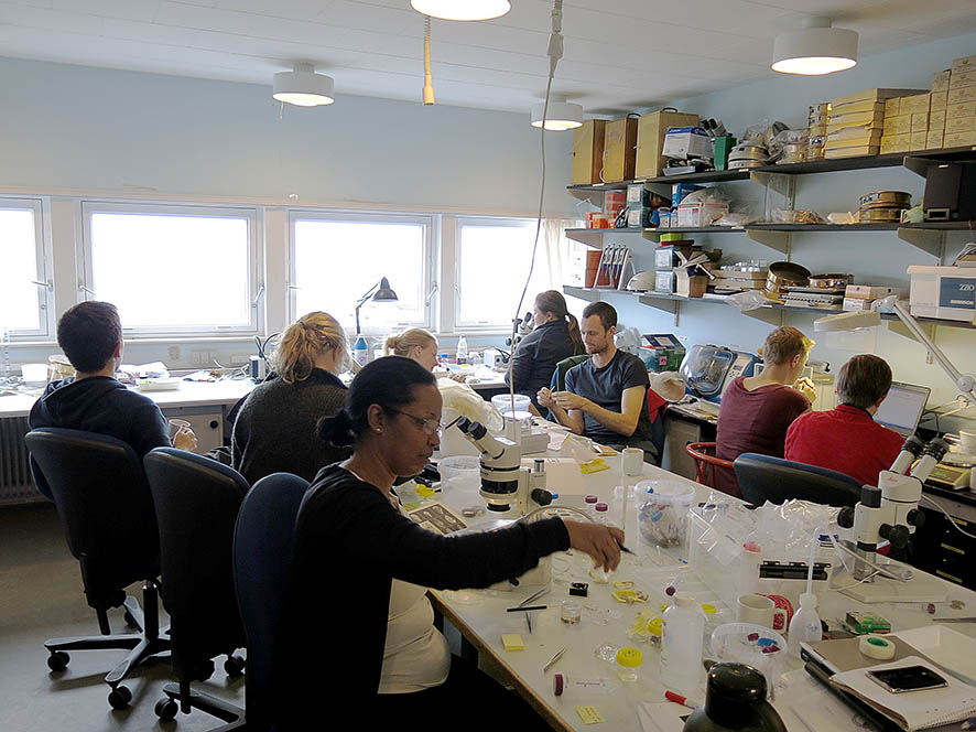
Photo: A Mucharin   Photo: A AltenburgerLast days in the lab: full house!                         The underwater film team: Mette and Jenny

Photo: I Meyer-Wachsmuth

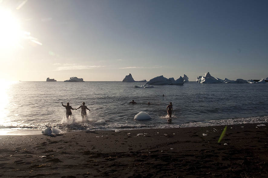
Bathing between icebergs – who can resist?

Photo: A Mucharin

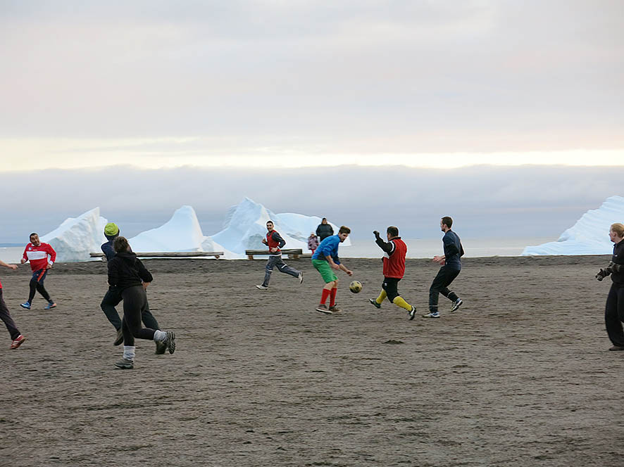
Football match against the Qeqertarsuaq “Old boys”, who turned out to be not that old… and pretty fit!

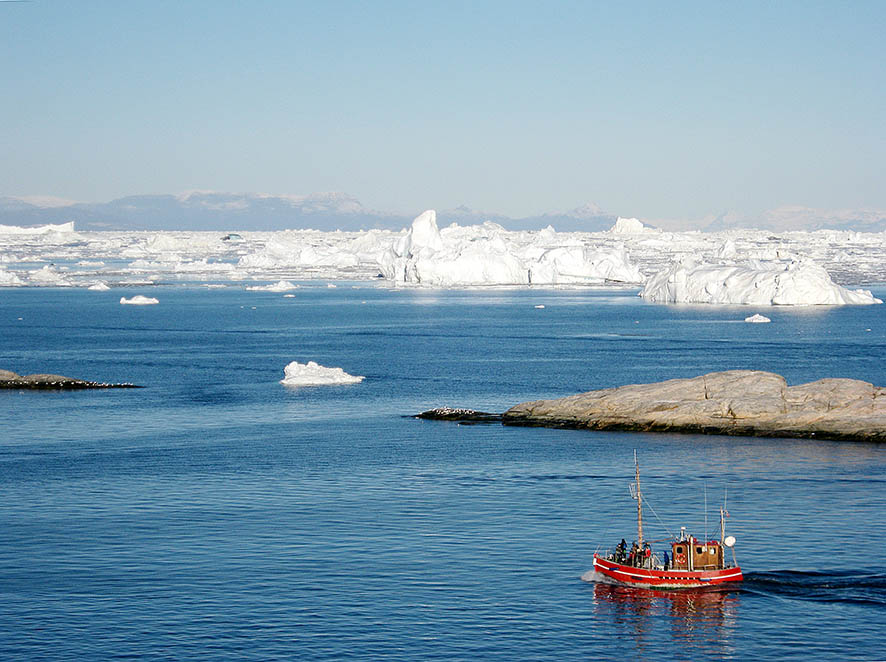
We left the Arctic station on a beautiful sunny day and headed towards Ilulissat, where we spent two days in wait for our flights back to first Kangerlussuaq and then Copenhagen. And beautiful days that was: Ilulissat is known for its icebergs and some of us took an icefjord tour on a handsome, oldish, boat with red paint and wooden deck. And – to our surprise – it turned to be out the old Porsild – the Arctic Station’s former research vessel!

Photo: D de Abreu

On the way to the ice fjord in Ilulissat – on board of the boat that turned out to be the Arctic Station’s former research vessel (the “old” Porsild!).

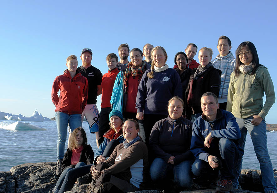
Now we are back to our respective homes – wrapping up coursework and getting on with our lives, PhD projects, master theses, scientific work and teaching. But we all agree: this was a very special course bringing us close to Arctic nature and providing us with outstanding possibilities to collect and study Arctic marine organisms.  We could both widen our taxonomic knowledge and – in different degrees – even get data that are of direct use for our ongoing research projects.
group-photo-beach-Peter-sm

At the end of this blog, we want to thank all those who helped us during this trip: Ole Stecher and Akaaraq Mølgaard at the Arctic Station, the crew of RV Porsild: skipper Frederik Grønvold and boatmen Søren and Johannes. Also, we are thankful to Reinhard Møberg Kristensen (Univ. Copenhagen) for suggestions concerning sampling sites and use of equipment!

logo-NorEng-colorNEW-small

The Research School in Biosystematics (ForBio) is funded by the Research Council of Norway and by the Norwegian Taxonomy Initiative – thanks for making a course like this possible!

And thanks to all of you who have been following us via this blog!

Written by: Christiane Todt (coordinator ForBio, University Museum of Bergen);                   Featured image: (Jenny) under the rainbow. Photo: Anne-Helene Tandberg


Photo: MHEilertsen & H Flørenes

Calcareous sponges of Disko Bay

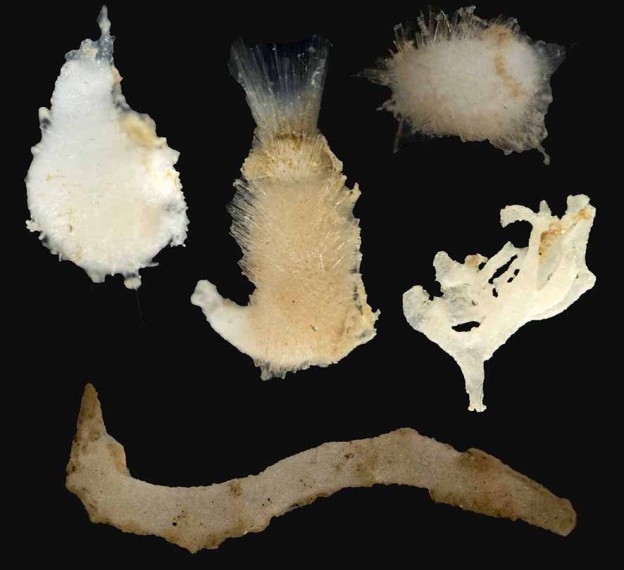
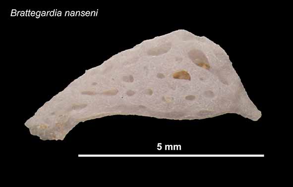
Fifteen species of calcareous sponges (phylym Porifera, class Calcarea) are known from the Disko Bay area, and nine of these have their type locality here, which means that this was the area they were fist described from. During this fieldtrip we wanted to collect calcareous sponges from Disko to get material that can be used for genetic studies to investigate the relationships between the populations in Disko Bay and populations of what is believed to be the same species in other localities.

Photo: MHEilertsen & H Flørenes

Photo: MHEilertsen & H Flørenes

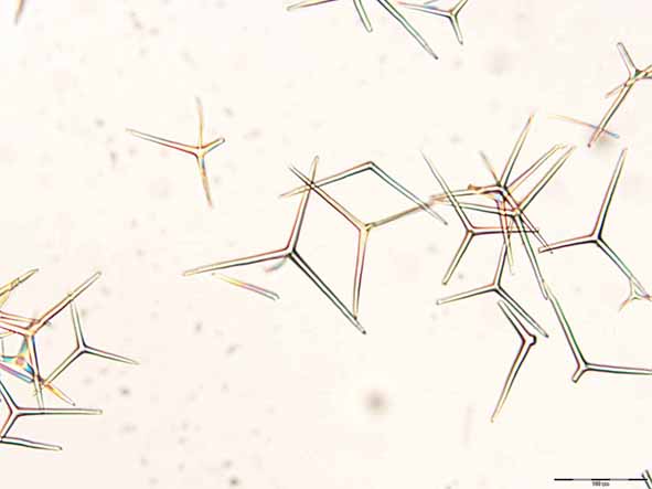
Spicules from Brattegardia nanseni.

Photo: MHEilertsen & H Flørenes

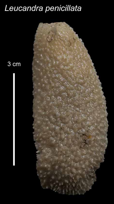
Largest calcareous sponge found during this fieldtrip.

Identifying calcareous sponges is not a straightforward business; first of all they are very small (most < 1 cm), and secondly they do not have any obvious external characters like bright colours, legs or even a head. In stead they have spicules. Spicules are small needle-like structures that function as the skeleton of the sponges, and in calcareous sponges they are made out of calcium carbonate. The shape of the spicules is an important character to identify sponges, in addition to the morphology of the sponge body and the arrangement of different types of cells. To look at the spicules of the sponges we need to clean the spicules from tissue and mount them on microscope slides. We cut out a piece of the sponge and add household bleach, which is left to work for 30 minutes, followed by three rounds of cleaning with distilled water, each taking 30 minutes. To make a permanent slide that we can look at again and again we use an imbedding medium that takes about 24 hours to dry sufficiently, so the time between we find a sponge specimen until we can identify it can be quite long.

Photo: MHEilertsen & H Flørenes

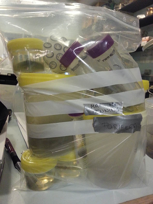
Collection of sponges fixated on ethanol.

We have had quite good sampling success so far, with over 50 specimens of calcareous sponges, and a few sponges of other classes. Most of the specimens were found sitting inside globular aggregations of Lithothamnion, a red algae (described in an earlier blogpost). Getting the sponges out from the Lithothamnion was tricky, both because they are very small and hard to spot, and because they were sitting quite far inside the globules, probably to get shelter. We also got many specimens from a triangular dredge sample that was sieved through a 1mm sieve. This is also where we found our largest specimen so far on this course! A beautiful Leucandra penicillata with a length of a whopping 5 cm! The calcareous sponges we have identified belong to at least eight different species, but the material has to be examined by experts to be certain of the identification.

Not all the samples we collect are equally interesting, but ones in a blue moon something special comes along. Yesterday, while sorting through the material from the dredge, we realized that we had just found the remnants of our good friend Spongebob Squarepants! It was a sad day indeed as we sieved out our global mascot from the murky waters. A memorial was thrown in his honour with the Greenlandic flag on half a pole. But life goes on and so does the search for new exciting sponges in different parts of the world!

Photo: Daniela de Abreu

Oh no – Sponge Bob is dead!!

By Henning and Mari (University of Bergen).

Featured image: Variety of calcareous sponges from Disko